|
|
|
|
Case Report |
Von
Meyenburg Complexes Associated with Polycystic Kidney Disease and The Endocystic Mural Nodule Sign: A Case Report Polikistik Böbrek Hastalığının Eşlik Ettiği Endokistik Mural Nodül Bulgusu
Olan Von Meyenburg
Kompleksi: Olgu Sunumu *Mehmet
TAHTABAŞI1 [ID], Ergin KARAMAN1 [ID], Emin ÇAKMAKCI2 [ID], Sadettin ER3 [ID] Abstract Von Meyenburg complexes (VMCs) are one of the rare, congenital, fibropolycystic benign diseases of the liver. We report a
40-year-old woman with characteristic imaging findings for diagnosis of VMCs
associated with polycystic kidney disease. Multiple micro-nodular lesions,
many of which were cystic, were detected on ultrasound (US). There were
echogenic mural nodules and comet tail artifacts in their internal
structures. In magnetic resonance imaging (MRI), the mural nodular
enhancement and starry sky appearance were observed. The presence of endocystic mural nodules in both US and MRI, as well as
the starry sky appearance that shows VMCs, are very important findings for
the diagnosis of this condition. Keywords:
Von Meyenburg complex, Bile
duct, Polycystic kidney, Endocystic nodule, Starry
sky appearance. Özet Von Meyenburg kompleksleri (VMC'ler),
karaciğerin nadir görülen doğuştan, fibropolikistik
iyi huylu hastalıklarından biridir. Polikistik böbrek hastalığı ile ilişkili VMC'lerin tanısı için karakteristik görüntüleme bulguları
olan 40 yaşında bir kadın olguyu sunuyoruz. Ultrasonografik (US)
görüntülemede çoğu kistik yapıda olan çok sayıda mikronodüler
lezyon tespit edildi. İç yapılarında ekojenik mural nodüller ve kuyruklu yıldız artefaktları vardı.
Manyetik rezonans görüntülemede (MRG), mural
nodüler kontrastlanma ve yıldızlı gökyüzü görünümü
gözlemlendi. Hem US hem de MRG'de endokistik mural nodüllerin
varlığı ve VMC'leri gösteren yıldızlı gökyüzü
görünümü bu durumun tanısı için çok önemli bulgulardır. Anahtar kelimeler: Von Meyenburg
kompleksi, Safra kanalı, Polikistik böbrek, Endokistik
nodül, Yıldızlı gökyüzü görünümü.
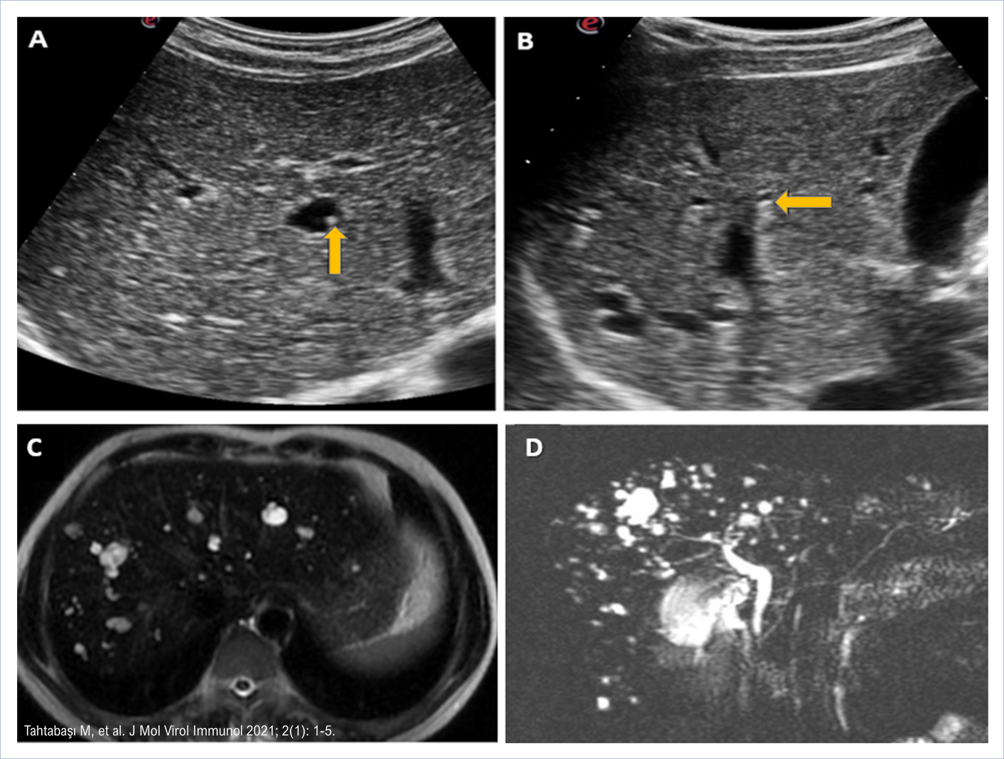
Figure 1. (A) The abdominal US showing the hyperechoic
mural nodule (arrow), and (B) echogenic foci with large posterior
comet-tail artifacts (arrow), (C) Axial T2-weighted and (D)
three-dimensional MRCP maximum intensity projection image showing a starry
sky appearance of well-circumscribed, multiple hyperintense lesions. Figure 1 png Şekil
1. (A)
Hiperekoik mural nodülü
gösteren abdominal US (ok) ve (B) Posteriorunda belirgin kuyruklu
yıldız artefaktları olan ekojenik odaklar (ok), (C)
Aksial T2- ağırlıklı ve (D) iyi sınırlı, çoklu
hiperintens lezyonların yıldızlı gökyüzü görünümünü gösteren üç boyutlu MRCP
maksimum yoğunluk projeksiyon görüntüsü. Şekil 1 png
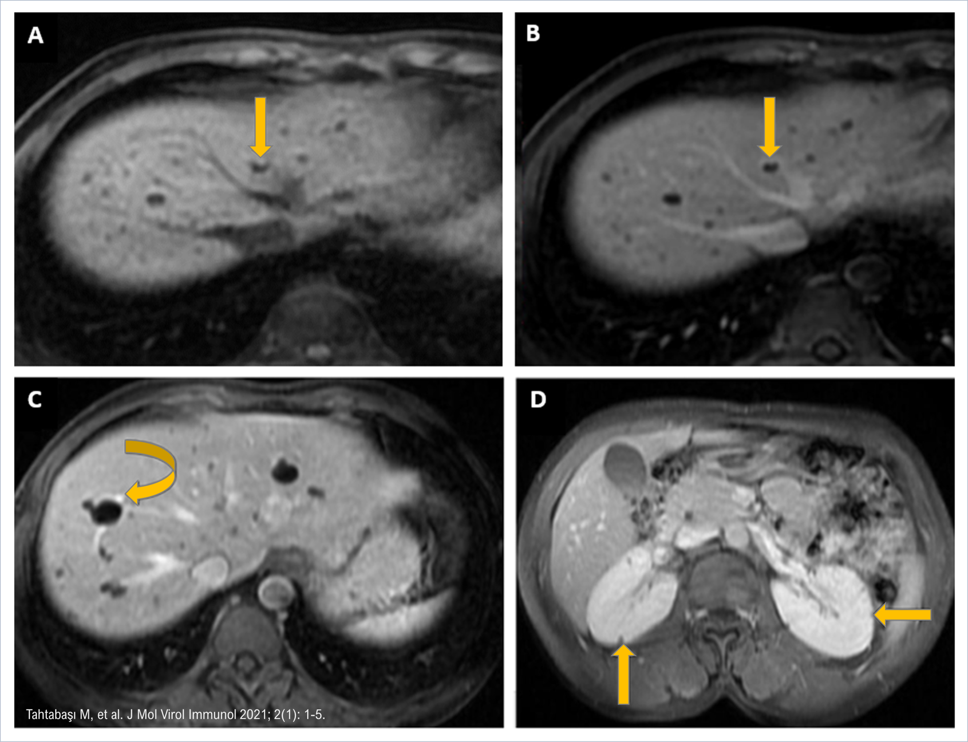
Figure 2. (A) T1-weighted MRI image showing multiple
low-signal lesions in the liver and the endocystic
polypoid projection of fibrous stroma (arrow), (B) gadolinium-enhanced
T1-weighted image demonstrating the enhancement of the mural nodule and
peripheral rim-like enhancement (arrow). (C) Post-gadolinium
T1-weighted image revealing peripheral enhancement of the lesions with
contrast filling in the portal vein adjacent to the lesions (curved arrow),
and (D) several microcysts (arrows) seen in the cortex of both kidneys. Figure 2 png Şekil
2. (A)
Karaciğerde çoklu düşük sinyalli lezyonları ve fibröz stromanın endokistik polipoid
projeksiyonunu gösteren T1 ağırlıklı MRG görüntüsü (ok), (B) mural
nodül ve periferik rim benzeri kontrastlanmayı
gösteren gadolinyum ile güçlendirilmiş T1 ağırlıklı görüntü (ok). (C) Lezyonların
bitişiğindeki portal vende kontrast dolgulu lezyonların periferik tutulumunu
gösteren post-gadolinyum T1 ağırlıklı görüntü (kavisli ok), ve (D) her iki böbreğin
korteksinde görülen birkaç mikrokist (oklar). Şekil 2 png |
|
DOI: 10.46683/jmvi.2021.23 |
|
|
Article in English |
|
|
|
|
|
|
|
|
1Department of Radiology, Mehmet Akif
Inan Education and Research Hospital, University of Health Sciences, Şanlıurfa, Türkiye. 2Department of Radiology, Ankara Dr.
Sami Ulus Education and Research Hospital, University of Health Sciences,
Ankara, Türkiye. 3Department of General Surgery, Ankara Numune Training and Research Hospital, University of
Health Sciences, Ankara, Türkiye. |
|
|
|
|
|
*Corresponding author Mehmet Tahtabaşı;
Asst.Prof., Department of Radiology, Mehmet Akif
Inan Education and Research Hospital, University of Health Sciences, Şanlıurfa, Türkiye. E-mail: mehmet.tahtabasi@sbu.edu.tr |
|
|
|
|
|
Received: 16.02.2021 Accepted: 01.03.2021 Published: 02.03.2021 |
|
|
Cite as: Tahtabaşı M, Karaman E, Çakmakcı
E, Er S. Von Meyenburg Complexes Associated with Polycystic Kidney Disease
and The Endocystic Mural Nodule Sign: A Case
Report. J Mol Virol Immunol 2021; 2(1): 1-5. |
|
|
|
|
|
View in academic indexes and databases |
|
|
|
|
|
|
|
|
|
|
|
|
|
|
|
|
|
|
|
|
|
|
|
|
|
|
|
|
|
|
|
|
|
|
|
|
|
|
|
|
|
|
|
|
|
|
|
|
|
|
|
|
|
|
|
|
|
|
|
Cited by 0 article*, 0 book chapter. |
|
|
|
|
|
©Copyright JMVI.
Licensed by Creative Commons Attribution-NonCommercial 4.0 International (CC
BY-NC 4.0). |
|